
A capillary refill check takes about five seconds and can decide whether a lip recovers fully or breaks down. It is also the test most often skipped, because pale, swollen lips after a dermal filler usually turn out to be harmless. Bruising is routine in the lips. Occlusion is rare, which is exactly why it gets missed.
This guide covers how ischaemia is recognised, how untreated tissue changes over hours and days, and how the current UK hyaluronidase protocol works, with the reasoning behind each step. It applies to cross-linked hyaluronic acid (HA) filler only. Hyaluronidase has no effect on non-HA products, so those cases need specialist advice immediately.
Signs of Vascular Occlusion After Filler: What Shows in the First Hour
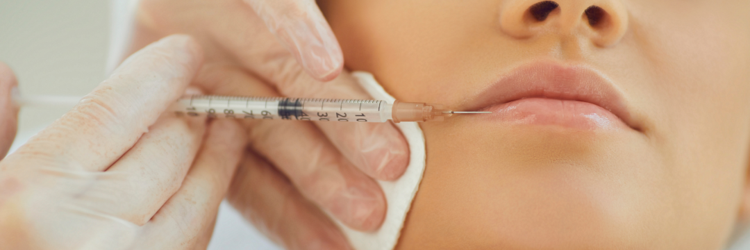
Most occlusions declare themselves during injection or within minutes. The CMAC guideline on filler-induced vascular occlusion describes ischaemic changes as typically instant or appearing within a few hours, so assessment starts before the needle leaves the tissue.
Pain that does not fit the procedure
Some discomfort is expected. Sudden escalating pain, or pain at a distant point such as the chin or nasal base, is not, and injection should stop at once. Two traps apply. Anaesthetic can mask pain until it wears off, and pain is sometimes absent in the early stages. A client who rings later that day with worsening pain needs a face-to-face review, not telephone reassurance.
Skin colour and capillary refill
Blocked arterial flow produces pallor first, then a purple net-like pattern called livedo reticularis as deoxygenated blood builds up in the venous plexus. The full territory of the artery needs checking, because filler and vasospasm can affect branches well away from the injection site.
Capillary refill is the key bedside test. Press until the skin blanches, hold for about five seconds, release, and time the return of colour. Under two seconds is normal. Compare both sides and note the client’s baseline before treatment. CMAC advises 30 to 45 minutes of observation after high-risk areas such as the nose, glabella and forehead. Where colour is uncertain after lip work, the same caution applies.
Bruise or Vascular Occlusion? Reading the Lip Correctly
Bruising is the usual finding after lip filler, so the working assumption is often wrong. The distinction rests on perfusion rather than colour. Bruised tissue has leaked blood but intact flow, so refill stays normal. Occluded tissue has blocked flow, so refill is slow or absent.
How the pattern differs
A bruise sits within tissue planes, often begins as pinpoint marks at needle entries and can drift downwards with gravity over hours. Occlusion follows a vascular territory, forms a branching or net-like pattern, can extend well beyond the treated area, feels cool and progresses instead of settling. Pain rises rather than eases. The two can coexist, so an obvious bruise does not exclude ischaemia beside it. A brisk refill on bluish skin points to venous compromise, which needs the same urgency.
Why the lip misleads
The superior and inferior labial arteries, both branches of the facial artery, supply the lips, and their position varies between individuals. CMAC therefore advises grading zones by risk and never describing any zone as safe.
Topical anaesthetic and the procedure itself can also cause blotchy pallor after topical anaesthetic, a frequent false alarm. If colour returns within two seconds inside the pale area, or both lips are equally blotchy (occluding both labial arteries at once is unlikely), anaesthetic is the likelier cause. Reassess at intervals rather than assuming either way.
Vascular Occlusion Stages and Why Time Matters
CMAC groups the skin changes of an untreated arterial occlusion into five stages. Pallor comes first, then reticulated purple mottling. Pustules typically appear around day three, the tissue darkens as haemorrhagic necrosis develops, and slough or eschar marks an open wound. Early stages can recover with minimal damage once flow returns. Later stages leave a wound that needs specific care.
Two mechanisms explain the urgency. Tissue tolerance depends on both the size and the duration of ischaemia, and thrombus forms early after intra-arterial HA. Reperfusion then adds injury of its own through free radicals and endothelial damage, and that injury grows with the length of ischaemia. Retinal tissue is far less forgiving. Older animal work suggested 97 minutes of tolerance, but CMAC cites recent research showing infarction within 12 to 15 minutes.

Vascular Occlusion Symptoms Confirmed: The Hyaluronidase Protocol
Once pain, pallor or delayed refill point to occlusion, treatment starts before the diagnosis is argued over.
Stop, tell the client, warm and massage
Injection stops immediately. The client is told plainly what is happening. Refill is compared on both sides, then firm massage with warmth encourages vasodilation and mechanical dispersal of the filler.
Preparing hyaluronidase
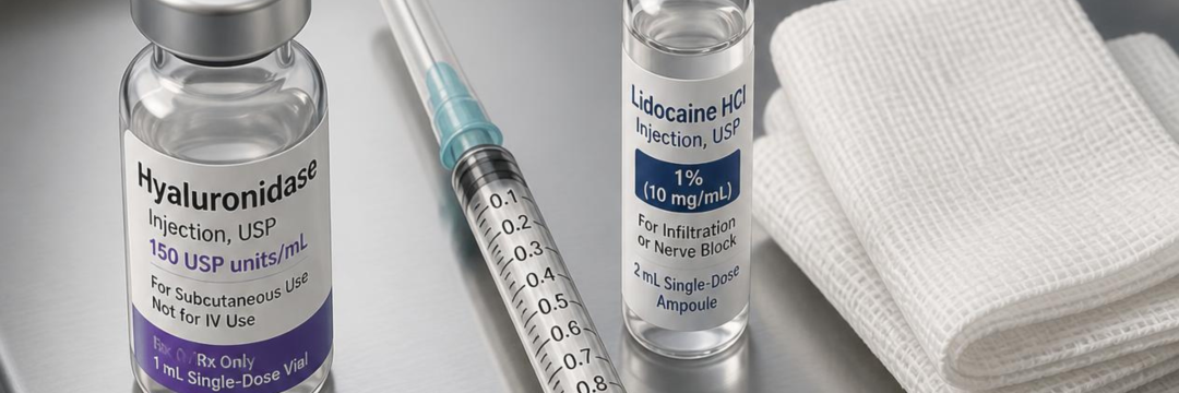
In the UK, hyaluronidase is a prescription-only medicine licensed to aid dispersal of injected fluids, so dissolving HA filler is off-licence and the client should be told this at consent. The CMAC hyaluronidase guideline sets storage below 25°C, immediate use of an opened ampoule and disposal of the remainder. It recommends reconstituting at 1,500 units per 1 mL where available, using lidocaine 1% or 2% without adrenaline. The anaesthetic makes treatment more tolerable and adds some vasodilation.
Non-prescribers need a protocol agreed with a prescriber in advance, and practitioners without one can find a prescriber. Stock levels are covered in this aesthetic emergency kit list.
Dose, area and re-dosing
The whole ischaemic area is treated, following the course of the artery, not only the injection point. Hyaluronidase diffuses across vessel walls after extravascular injection, so it does not need to enter the artery.
Two schedules exist. The ACE Group hyaluronidase guideline advises infiltrating 450 to 1,500 units over the area, reassessing at 60 minutes and repeating hourly for up to four cycles. The later modified high-dose hyaluronidase protocol from CMAC gives the initial dose, firm massage and reassessment of flow, then redosing at around 15 to 20 minutes if perfusion has not returned. The reasoning is pharmacological: rodent data give a tissue half-life of about five minutes after subcutaneous injection, which makes hourly gaps look long.
Where ultrasound is available, CMAC cites series clearing emboli with an average of 35 to 60 units, and up to 150 in a few cases. Treatment continues until refill and colour normalise.
Adjuncts and what to leave out
CMAC considers aspirin 300 mg stat, then 75 mg daily until reperfusion, reasonable if safe, though direct evidence is lacking. Clopidogrel is the alternative in aspirin allergy. It advises against GTN paste, because dilating vessels may push filler into smaller arterioles, and against sildenafil. Steroids are avoided routinely as they can impair wound healing, and oral antivirals are reserved for tissue that has begun to break down.
Allergy and skin testing
CMAC does not recommend skin tests in an emergency. Anaphylaxis is very rare, with only four published case reports since 1949 of reactions needing adrenaline, and no validated test concentration exists. Adrenaline must be in the kit. A history of bee or wasp sting allergy should be asked about, because venom contains hyaluronidase. Managing a reaction is covered in anaphylaxis in clinic.
When vision or neurology is involved
Visual disturbance, eye pain, ptosis or stroke-like features change the pathway. The UK consensus guidelines on filler-induced vision loss direct the client to the nearest emergency department without delay. Best practice is for the practitioner to accompany them, carrying at least 7,500 units of hyaluronidase.
Timolol 0.5% (one drop), a single 300 mg aspirin, paper-bag rebreathing and ocular massage are acceptable first steps only if they do not delay transfer. About 20 percent of cases involve the central nervous system, which is why the main emergency department is preferred. Practitioners who want a clinician on the line during any of this can call the Complications Consultant service.
Delayed Vascular Occlusion Filler Presentations and Follow-Up
Why onset can be late
Ischaemia usually appears at once or within hours, but CMAC describes onset days later and reports more clients arriving at a later stage. Three explanations are proposed. An embolus may lodge in a narrower distal arteriole. HA may absorb water and enlarge the bolus until it compresses or blocks a vessel. Or platelet aggregation may gradually complete an initial partial occlusion.
Treating late presenters
Hyaluronidase is still given, even where necrosis is established, because dissolving the embolus can protect tissue that remains viable. Late tissue often shows several stages at once. Once the skin breaks down or blisters, at roughly 72 hours, wound support and infection prevention take priority, with antibiotics only when clinically indicated.
Follow-up and records
Every client leaves with written warning signs and an emergency number, which an aftercare form can carry. A resolved occlusion still needs review the next day, and a wound needs follow-up until healed.
Photographs and contemporaneous notes matter. CMAC advises informing the insurer early because these events can lead to claims, and practitioners reviewing cover can compare aesthetics insurance options. Suspected product-related events also belong in Yellow Card reporting for aesthetic practitioners. The digital consent form should already cover off-licence hyaluronidase use and, for higher-risk areas, the rare risk of vision loss and stroke.
Before the next filler list, save the Complications Consultant helpline number where the whole team can find it. It costs £9 per month through Faces and connects practitioners to a medical professional by phone, email or video at any hour, so a suspected occlusion at 9pm on a Saturday does not have to be managed alone.
FAQs
How long after filler can a vascular occlusion appear?
Usually within minutes to a few hours of injection. Rarely, changes appear days later. Any new pain, blanching or mottling after treatment warrants same-day face-to-face assessment, whatever the interval.
What does a vascular occlusion look like on the lips?
Pallor, then purple net-like mottling, often with a cool feel and pain that keeps rising. Capillary refill is slower than two seconds compared with the untreated side. The change can extend beyond the lip along the arterial path.
How much hyaluronidase is needed for a vascular occlusion?
There is no single figure. Guidance ranges from 450 to 1,500 units over the affected area, repeated until flow returns. CMAC reconstitutes at 1,500 units per 1 mL and redoses after 15 to 20 minutes if perfusion has not returned. Ultrasound guidance can reduce the units needed.
Is a skin test needed before hyaluronidase in an emergency?
No. CMAC advises against skin testing in a suspected occlusion because anaphylaxis risk is minimal and no validated test concentration exists. Adrenaline should be available, and the client’s allergy history, including bee and wasp stings, checked.
What should happen if a client reports changes in vision?
Treat it as a medical emergency. The client goes to the nearest emergency department without delay, accompanied by the practitioner with at least 7,500 units of hyaluronidase where possible. First-aid measures must not delay transfer.