
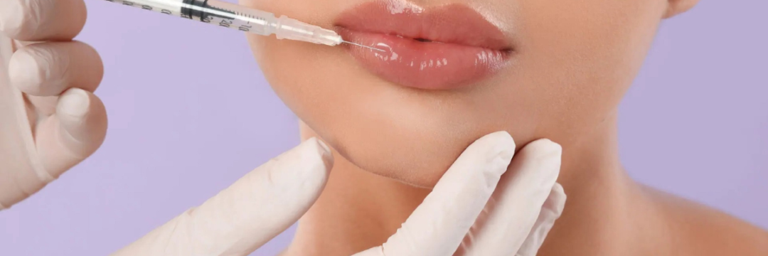
Not every lump after lip filler is the same problem, even though most patients, and plenty of practitioners, describe them all the same way: “a nodule.” One firm bump might clear on its own within a week. Another might need hyaluronidase and a steroid. A third might need weeks of antibiotics because it’s a biofilm the immune system can’t get near. The physical finding looks identical at first glance. What separates them is timing, and that’s exactly why an accurate digital consent form recording what was injected, where and when, alongside a properly logged aftercare form noting how the tissue looked at review, often does more diagnostic work than the physical exam itself.
Surface Lip Filler Nodules: Non-Inflammatory and Usually Early
Not every lump after filler is the same clinical problem. The first branch point in any work-up is timing. Nodules that appear within hours or days of treatment, are non-tender, non-erythematous and palpable close to the skin surface are usually non-inflammatory. They tend to result from uneven product distribution, injection that was placed too superficially, or too large a volume deposited in one point rather than spread across the treatment plane. In the lip specifically, this is more likely at the vermillion border and philtrum columns, where the tissue is thin and product placed even slightly too shallow becomes palpable, sometimes visible, immediately.
These early, superficial nodules typically respond to gentle massage in the first 48 to 72 hours, and to hyaluronidase where massage alone hasn’t resolved them within a week or two. They are not usually a sign of infection or immune reaction, and reassurance plus a short review interval is often all that’s needed before considering dissolving the area.
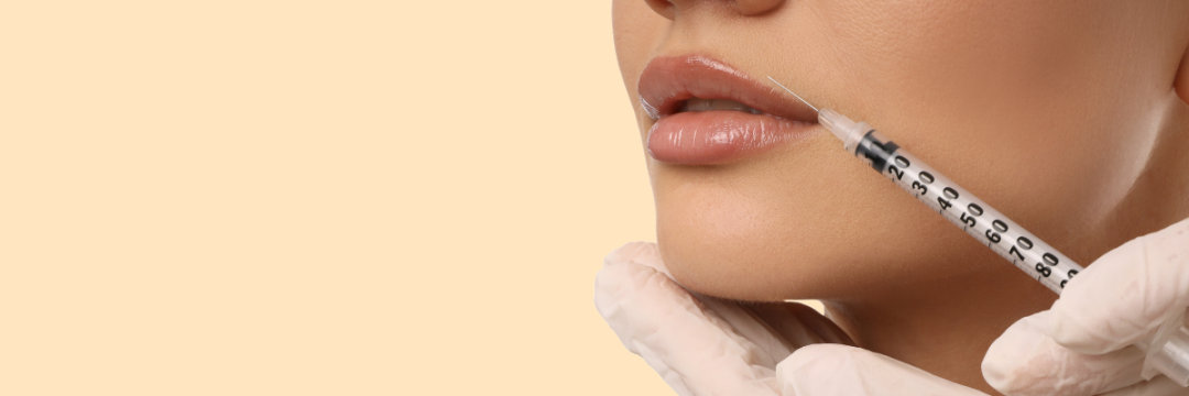
Delayed Onset Nodules Filler: When the Timeline Points to Biofilm or Immune Response
The clinical picture changes once a nodule appears after a genuine quiet period, typically defined in the literature as at least two weeks after treatment, sometimes months or years later. This delayed-onset pattern is what should prompt a broader work-up rather than a default to massage and reassurance.
Three broad mechanisms are usually considered for a delayed nodule: a biofilm, or low-grade bacterial colonisation on the filler surface that evades routine detection; a delayed-type hypersensitivity reaction driven by T-cell activity against the filler material; and foreign body granuloma formation, where the immune system walls off filler it cannot clear. A useful diagnostic principle described in a review of making sense of late tissue nodules associated with hyaluronic acid injections is that a period of genuine quiescence, tissue looking and feeling normal for days, weeks or months before the nodule appears, helps rule out a simple post-procedural response such as residual bruising or swelling, which can otherwise be mistaken for a true delayed reaction.
History-taking should cover any intercurrent illness, dental work, other injectable treatments (including in different areas, since delayed nodules can be triggered by a subsequent unrelated procedure), and any recent vaccination, all of which have been described as potential triggers for a previously quiet filler site to flare.
Cheek Filler Nodules vs Lip Filler Nodules: Does Location Change the Work-Up?
Anatomically, the work-up principles are the same for cheek filler nodules and lip nodules: timing, inflammatory signs, fluctuance and response to a quiet period all matter more than the treatment area itself. What differs is the differential diagnosis practitioners need to keep in mind. In the cheek, a firm mass should also prompt consideration of a reaction near the parotid duct o a misplaced deep bolus close to vascular structures, since a slow-growing lump in this region can, rarely, mimic early signs of vascular compromise. Distinguishing a nodule from a genuine vascular event matters clinically: a nodule is typically painless or mildly tender, doesn’t blanch abnormally, and the surrounding skin colour and capillary refill are normal, whereas vascular compromise presents very differently and needs immediate management rather than a staged work-up. Practitioners who want a structured way to rule this out quickly can review the capillary refill and skin-mottling checks covered in recognising and reversing vascular occlusion in lip filler.
In the lip, the main differential to exclude first is a simple non-inflammatory nodule from technique, as covered above, before moving on to the delayed-onset pathway.
Granuloma Lip Filler Nodules: Diagnosis and Management
A granuloma lip filler nodules presentation is one of the more disfiguring and persistent delayed reactions, and one of the most commonly mislabelled. A true foreign body granuloma is a chronic inflammatory response to a foreign material the body cannot phagocytose or break down, and it typically appears months after treatment, is cystic or firm, may have a purplish hue from congested dermal capillaries, and often affects multiple injection sites rather than just one. Estimated incidence for HA fillers sits at roughly 0.02 to 0.4 percent, low, but not negligible given the volume of lip filler procedures performed each year.
The important clinical caveat is that a definitive granuloma diagnosis technically requires histological confirmation. In practice, most patients are unwilling to undergo biopsy for a cosmetic complication, so the accepted approach, reflected in the proposed treatment algorithm for delayed inflammatory reactions to hyaluronic acid fillers, is to work through management in a stepwise way rather than waiting for tissue confirmation: intralesional hyaluronidase first where the filler is HA-based, intralesional corticosteroid if hyaluronidase alone doesn’t resolve it, and a swab or aspirate for culture and PCR if there’s any suspicion of biofilm or atypical infection, since biofilms frequently fail to grow on standard culture and need a longer, often combined antibiotic course, typically several weeks, alongside hyaluronidase to help break down the protective matrix around the bacteria. Surgical excision and intralesional 5-fluorouracil are reserved for cases that don’t respond to this staged approach.
Granulomas Meaning in an Aesthetic Context
Outside dermal filler medicine, granulomas meaning is broader: a granuloma is simply a small area of chronic inflammation, made up of macrophages and other immune cells, that forms when the body encounters material it can’t clear quickly, whether that’s a foreign body, certain infections, or an autoimmune process. In aesthetics, filler-associated granulomas are one specific example of this general mechanism, which is why an aesthetic practitioner’s differential for any longstanding nodule should always include causes that have nothing to do with the injection at all.
Skin Granulomas Beyond Filler: Other Causes to Rule Out
Before attributing every firm lump to the last filler treatment, it’s worth remembering that skin granulomas have causes unrelated to cosmetic injectables: retained suture material, insect bites, tattoo pigment reactions, and systemic granulomatous conditions such as sarcoidosis can all present as a firm dermal nodule. A history that doesn’t fit the expected filler timeline, multiple nodules appearing in areas that have never been treated, or systemic symptoms such as fatigue, joint pain or eye involvement should prompt referral rather than continued in-clinic management, since these point toward a cause the aesthetic clinic isn’t equipped to investigate or treat.
This is also where documentation earns its keep. A clear record of exactly what was injected, where, and when, cross-referenced against when the nodule first appeared, is often what separates a straightforward filler-related work-up from a referral that saves the patient months of the wrong treatment.

Start Training With Faces Training Academy
Recognising which pathway a nodule falls into, non-inflammatory, delayed hypersensitivity, biofilm or true granuloma, takes more than reading a single article. It’s a skill built through supervised case exposure. Practitioners who want structured, hands-on teaching in complications recognition and management, rather than picking it up reactively in clinic, can find advanced injectables and complications-focused courses through Faces Training Academy, delivered in small groups with expert supervision.
Talk to a Complications Consultant Before You Guess
Practitioners who see a lump they’re not confident diagnosing should log the case properly rather than guess. A Complications Consultant can talk through the presentation and next steps before treatment decisions are made.
FAQs
Do filler nodules go away?
Many do, particularly early, non-inflammatory nodules, which often resolve within days to a couple of weeks with massage or a single round of hyaluronidase. Delayed, inflammatory nodules and true granulomas are less predictable and can persist for months without targeted treatment, so ongoing swelling shouldn’t simply be watched and waited on indefinitely.
What are granulomas?
A granuloma is a small pocket of chronic inflammation that forms when the immune system encases material it cannot break down or clear, most often immune cells clustered around a foreign body, and in aesthetics this is usually filler material. They tend to develop months after treatment, can affect more than one injection site, and generally need active management rather than simply resolving on their own.
How do you tell if a filler lump is infected or just inflamed?
A true infection usually comes with warmth, spreading redness, pain out of proportion to the size of the lump, and sometimes fever, whereas an inflamed nodule from a delayed hypersensitivity reaction tends to be firm, mildly tender at most, and doesn’t spread. When it’s genuinely unclear, a swab or aspirate for culture and PCR is more reliable than a visual judgement, since biofilm-driven cases can look deceptively calm on the surface while still needing an extended antibiotic course.
Does hyaluronidase get rid of a granuloma?
Hyaluronidase can help where the granuloma still contains identifiable HA filler, since dissolving the material removes what the immune system is reacting to. It’s less reliable once the reaction has become more fibrotic or the material has been walled off for a long period, which is why intralesional steroid, and occasionally surgical excision, are added as next steps rather than repeating hyaluronidase indefinitely.
How long should you wait before treating a delayed-onset nodule?
There’s no fixed waiting period, but most protocols support starting assessment as soon as a nodule has been present for more than a few days without settling, rather than defaulting to a long observation window. Earlier intervention, once infection has been reasonably excluded, tends to produce a faster resolution than letting a nodule sit for weeks on the assumption it will clear on its own.